발간보고서
home > 자료실> 발간보고서
- 글자크기
Types of cancer and it’s classification
| 작성자 | 관리자 | 카테고리 | 전문가 인사이트 |
|---|---|---|---|
| 작성일 | 2018-12-18 | 조회수 | 3,239 |
| 원문 | |||
| 출처 | |||
한국보건산업진흥원 해외제약전문가 이민영 박사
Types of cancer and it’s classification
[Reference: American Joint Committee on Cancer, www.cancerstaging.org]
Cancer is mainly categorized according to the main organ in which the cancer started in, and the type of organization and tissue. Then, cancer is further classified into stages and grades according to the evaluation systems that describe the extent of cancer. The most appropriate options to treat with a cancer will be decided based upon the cancer type and its classifications identified.
To confirm the cancer type and its classifications, patients may undergo radiological examinations such as CT scans (computed tomography), MRI (magnetic resonance imaging) or PET scans (positron emission tomography), and even gene mutation tests.
Staging of cancer
An important point before starting to treat with cancer is to first find out how far it has spread or what “stage” it has reached. Staging is a system that is used to classify the extent of cancer and to decide what treatment option would be the best for patients.
Usually, your doctor tells the stage of cancer at the time of diagnosis and further finds out the “clinical stage” of the disease. A pathologist will exam the tumor tissue and assign a “pathologic stage”. In general, the pathologic stage is the most important information in making treatment decisions.
What is the TNM system?
Doctors use the tumor, lymph node and metastasis (TNM) system for staging most cancers. The American Joint Committee on Cancer (AJCC) developed the TNM system, maintained and updated it. It is the standard system for cancer staging used by physicians and scientists.
-
The ‘T’ refers to tumor, which indicates tumor size, extent or penetration.
-
The ‘N’ stands for node, which indicates the number of lymph nodes with cancer or the location of the cancer-involved lymph nodes.
-
The ‘M’ stands for distant metastasis or the spread of the cancer to other parts of the body. It indicates that there are cancer cells outside the local area of the tumor and its surrounding lymph nodes.
The most common cancers that doctors stage using the TNM system are breast, colon, renal, stomach, esophagus, pancreas, and lung cancers. Other cancers staged with the TNM system include soft tissue sarcoma and melanoma. Staging systems exist for 52 sites or types of cancer.
Some cancers are not staged using the TNM system. Cancers of the blood, bone marrow, brain and some other sites might not use the TNM staging. Gynecologic cancers use another staging system which doctors can translate into TNM.
TNM staging:
-
T (Tumor): tumor size, extent or penetration
-
N (Nodes): the number of lymph nodes with cancer or the location of the cancer-involved lymph nodes
-
M (Metastasis): distant metastasis or spread of the cancer to other parts of the body, indicating cancer cells outside the local area of the tumor and its surrounding lymph nodes
T: size or direct extent of the primary tumor
-
Tx: tumor cannot be evaluated
-
Tis: carcinoma in situ, is a group of abnormal cells or pre-cancer cells
-
T0: no signs of tumor
-
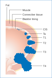
T1, T2, T3, T4: size and/or extent of the primary tumor, classified according to tumor size and involvement in surrounding tissues
[Diagram showing the T stages of bladder cancer]
N: degree of spread to regional lymph nodes
-
Nx: lymph nodes cannot be evaluated
-
N1: tumor cells present in regional lymph nodes (at some sites: tumor spread to closest or small number of regional lymph nodes)
-
N2: tumor spread to an extent between N1 and N3 (N2 is not used at all sites)
-
N3: tumor spread to more distant or numerous regional lymph nodes (N3 is not used at all sites)
M: presence of distant metastasis
-
M0: no distant metastasis
-
M1: metastasis to distant organs (spread beyond regional lymph nodes)
Overall stage grouping
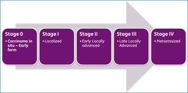
Overall Stage Grouping is referred to as Roman Numeral Staging to describe the progression of cancer.
-
Stage 0: carcinoma in situ, the cancer is confined to the epidermal cells only.
-
Stage I: cancers are localized to one part of the body. The original tumor is small and confined within the organ it started in. àStage I cancer can be surgically removed if small enough.
-
Stage II: cancers are locally advanced and has spread to lymph nodes close to the tumor. à Stage II cancer can be treated by chemo, radiation or surgery.
-
Stage III: cancers are also locally advanced and has spread to lymph nodes and surrounding tissues. The specific criteria for Stages II and III differ according to diagnosis. à Stage III can be treated by chemo, radiation or surgery.
-
Stage IV: cancers have often metastasized, spread to other organs or throughout the body. à Stage IV cancer can be treated by chemo, radiation or surgery.
What does ‘stage grouping’ mean?
Once doctors have determined the TNM categories, they can classify the cancer into a ‘stage group’. Stage grouping uses Roman numerals I, II, III, or IV. The larger the number, the more advanced the stage of cancer will be.
If you will take a part in a clinical trial, the stage group of the cancer must be known first. It will allow you to be placed into a proper treatment group. (A clinical trial is a research study that is conducted, with the patient’s permission to investigate how effective and safe a new drug treatment is.)
That means, staging is a system that is used to classify the extent of cancer and to decide what treatment option would be the best for patients.
Grades of cancer cells
Grading in cancer is different from staging. Staging is a measure of the extent to which the cancer has spread. Grading is a measure of the cell appearance in tumors.
Cancer grade is the description of how abnormal the cancer cells look like under a microscope when compared to the surrounding normal tissue. The grades are given from 1 to 4.
-
Grade 1 – well differentiated cells, looking quite similar to surrounding normal cells with slightly unusual forms of separation (low grade à means slow growing cells)
-
Grade 2 – moderately differentiated cells, with more unusual forms compared to Grade 1 (intermediate grade)
-
Grade 3 – poorly differentiated with unusual forms. Cancer cells are abnormal looking and lack normal tissue structure (high grade à tend to growing rapidly and spread fast)
-
Grade 4 – Undifferentiated. Cancer cells are immature and cannot be distinguish with primitive form (high grade à tend to growing rapidly and spread fast)
Types of cancer based on the origin of organ or tissue
-
Carcinoma
-
Sarcoma
-
Myeloma
-
Leukemia
-
Lymphoma
-
Mixed Types
Carcinoma
Carcinoma is a type of cancer that develops from epithelial cells, or in tissues that line the inner or outer surfaces of the body. This cancer accounts for 80-90% of all cancers.
Sarcoma
Examples of sarcoma are malignant tumors made of cancellous bone, cartilage,fat, muscle,vascularor hematopoietictissues. Sarcoma is different from carcinoma, which originates from epithelial cells.
Myeloma
Known as multiple myeloma or plasma cell myeloma, myeloma is a cancerof the plasma cells, a type of white blood cells that are normally responsible for producing antibodies.
Leukemia
Leukemia is a group of cancers that usually begins in the bone marrow and result in high numbers of abnormal white blood cells. These white blood cells are not fully developed and are called blastsor leukemia cells. Leukemia can be further broken down into the followings:
-
Myelogenous or granulocytic leukemia: cancerous change that takes place in marrow cells, which form red blood cells, some white blood cells and platelets.
-
Lymphoblastic leukemia: cancerous change that takes place in a type of marrow cells, which form lymphocytes that are infection-fighting immune system cells
-
Polycythemia vera or erythremia: bone marrow disease that leads to an abnormal increase in the number of blood cells, especially red blood cells.
|
Cell type |
Acute |
Chronic |
|
Lymphocytic leukemia |
||
|
Myelogenous leukemia |
Acute myelogenous leukemia |
Lymphoma
Lymphoma is a cancer that originates in infection-fighting cells of the immune system, called lymphocytes. These cells are found in the lymph nodes, spleen, tonsils, thymus and other parts of the body. Lymphoma is different from leukemia – lymphoma starts in infection fighting cells, while leukemia starts with blood forming cells inside the bone marrow.
The two main categories of lymphoma are: (1) Hodgkin’s lymphoma, and (2) Non-Hodgkin’s lymphoma. Hodgkin’s lymphoma is marked by the presence of a type of cells called Reed-Sternberg cells, while all other lymphomas are categorized as Non-Hodgkin’s lymphomas.
Mixed Types
Mixed types of cancers in the variety of tissues or organs. Some examples are:
-
Adenosquamous carcinoma
-
Mixed mesodermal tumor
-
Carcinosarcoma
-
Teratocarcinoma
Classification by Main Organ that Cancer started in
Cancer is used to be classified according to the organ it started in. For example: lung cancer, stomach cancer, colorectal cancer, pancreatic cancer, liver cancer, brain cancer, head & neck cancer, oral cancer, skin cancer, kidney cancer, bladder cancer, prostate cancer, breast cancer, ovarian cancer, etc.
Which category or classification is my cancer belong to? What kind of genetic mutations occurred in my cancer?
There are various cancer types. They are categorized according to the main organ in which cancer started in and the type of organ and tissue. As discussed, cancer is classified into stages and grades according to the evaluation of TNM staging system and pathological grading of cancer cells, respectively.
To do accurate evaluation of the stage and grade of cancers, doctors can make use of scan imaging tools. Nowadays, doctors can also test and determine the specific gene mutations that caused the cancer, for example, HER2, RAS, BRAF, KRAS, EGFR, ALK, p53, etc., though the technology of gene mutation test is rapidly evolving but it is not feasible yet to cover all cancer types at this moment.
These kinds of modern technology are useful to evaluate the extent of cancer and to help us decide on the best treatment options for cancer patients - for surgery, chemotherapy, radiotherapy or a combination of treatment, target therapy, and immunotherapy, etc.


